
Plasmon Coupling
What is plasmon coupling and why does it occur?
Surface plasmons are a collective free electron density oscillation on the surface of metals in response to an external electric field (1,2). At large interparticle distances, the surface plasmon resonance is based on the size and geometry of the nanoparticle. However, at close proximity plasmon coupling can occur, where the surface plasmons of adjacent nanoparticles interact.
This coupled (3), or coherent oscillation of electrons across the surface of two or more particles in close contact, causes the optical properties of the coupled particles to differ from that of an individual particle in solution.
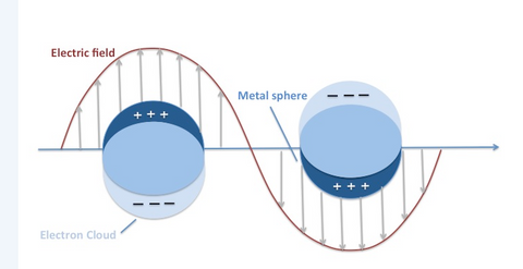
Figure 1: Electron Delocalization. A surface plasmon is characterized as a surface charge density wave at the metal surface
Factors influencing plasmon coupling
The optical effects of plasmon coupling can vary vastly, depending on nanoparticle shape and size, interparticle spacing, number of plasmon coupled nanoparticles, light polarization and other factors. For example, plasmon coupling of gold nanospheres results in a significant shift of the overall extinction peak (dominated by absoption) to higher wavelengths as the interparticle spacing is reduced (Fig. 2).
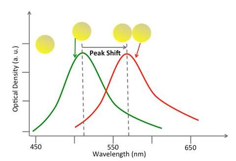
Figure 2: Single gold nanosphere absorbance spectrum in green, plasmon coupling absorbance spectrum in red.
Related applications
The plasmon coupling effect can be used in biological applications to detect the presence of certain cell types and image them non-invasively. Gold nanospheres can be conjugated to antibodies that specifically target surface receptors on cancer cells. When these targeted gold nanospheres bind to cancer cells, they are engulfed into the cell via a process called receptor-mediated endocytosis. After cellular uptake, the nanospheres are confined in high concentrations inside of vesicles called endosomes. The tight packing of the particles in endosomes leads to plasmon coupling.
Interestingly, the optical spectral shifts which accompany plasmon coupling can be detected using imaging techniques such as photoacoustic imaging. Thus, photoacoustic imaging of gold nanospheres delivered to tumors can be used to non-invasively locate cancer cells inside tissue. This specific use of plasmon coupling has been employed to detect metastasis in sentinel lymph nodes and image primary tumors (4-6).
Silica coating can mitigate plasmon coupling effects
In some applications using plasmonic nanoparticles as contrast agents, such as gold nanoparticles, plasmon coupling is not always desired since it can change the optical spectra (7), which may be critical for imaging modalities with limited optical bandwidth.
Plasmon coupling can be avoided by silica coating the nanoparticle to ensure interparticle distances are sufficiently far. Even under high packing conditions found in endosomes, a silica layer of sufficient thickness can eliminate plasmon coupling (7). Furthermore, silica is transparent in the visible and near infrared wavelengths, and does not significantly alter the optical properties of the core plasmonic nanoparticle. Silica coating has also been shown to increase photoacoustic imaging signal in comparison to uncoated particles (8).
Is the plasmon coupling effect helpful? Depends.
Plasmon coupling can hinder some applications (e.g., contrast-guided imaging with gold nanoparticles) or can be exploited to enable detection of physiological events or enhance the detection of small molecules (e.g., surface enhanced Raman spectroscopy or SERS).
Contact us for more information about the plasmonic properties of gold nanoparticles. We would be glad to help you figure out if our particles are a good fit for your research.

References:
- Surface plasmon resonance in nanostructured metal films under the Kretschmann configuration. Leong et al, J. Applied Physics 106 (2009).
- Localized Surface Plasmon Resonance Spectroscopy and Sensing. Willets et.al, Annual Review of Physical Chemistry 58 (2007).
- Plasmonic coupling in noble metal nanostructures. Jain et.al, Chemical Physics Letters 487, 4-6 (2010).
- Sentinel lymph node biopsy revisited: ultrasound-guided photoacoustic detection of micrometastases using molecularly targeted plasmonic nanosensors. Luke et al, Cancer Research 74-19 (2014).
- Multiwavelength photoacoustic imaging and plasmon resonance coupling of gold nanoparticles for selective detection of cancer. Mallidi et al, Nano Letters 9-8 (2009).
- Visualization of molecular composition and functionality of cancer cells using nanoparticle-augmented ultrasound-guided photoacoustics. Mallidi et al, Photoacoustics 3-1 (2015).
- Preventing Plasmon Coupling between Gold Nanorods Improves the Sensitivity of Photoacoustic Detection of Labeled Stem Cells in Vivo. Comenge et al, ACS Nano 10-7 (2016)
- Silica-Coated Gold Nanorods as Photoacoustic Signal Nanoamplifiers. Chen et al, Nano Letters 11-2 (2011).
Related links:
Plasmonics 101: What is surface plasmon resonance?