 Applications
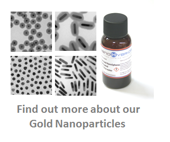
Our gold nanoparticles are excellent for providing contrast in a wide variety of biological applications.

Photoacoustic (Optoacoustic) Imaging
Photoacoustic Imaging technology is a hybrid optical imaging technique that combines optics and acoustics. In photoacoustic imaging, the tissue is illuminated with a laser light of a specific wavelength. This causes the tissue to absorb light and heat up which in turn leads to thermoelastic expansion emitting an acoustic wave. This wave is detected by an ultrasound transducer to generate an image.
Contrast agents such as gold nanoparticles can highly enhance both the sensitivity and specificity of photoacoustic imaging. Find out more about photoacoustic imaging and its advantages over other in vivo imaging modalities.
Optical Coherence Tomography (OCT)
OCT, another optical imaging technique, employs near-infrared light to penetrate into scattering media, like biological tissue, and generate 3D images using an interferometric techinique.
OCT has broad applications in diagnostic medicine, notably in ophthalmology and interventional cardiology. Our nanoparticles, especially our nanorods that scatter and absorb near-infrared light are excellent contrast agents for OCT, allowing researchers and scientists to not just visualize anatomy, but to probe molecular targets in diseased tissue.
Multiphoton Microscopy – Two-Photon Luminescence (TPL) and Second-Harmonic Generation (SHG)
Multiphoton microscopy is an optical imaging technique that is known for its ability to generate high resolution images up to depths of 1 millimeter in biological samples.
A high flux of excitation photons is required for sample illumination, typically from a femtosecond laser source. Pulsed laser sources, like femtosecond lasers used for multiphoton microscopy and nanosecond lasers used in photoacoustic imaging, deliver large payloads of energy over extremely short times.
This type of excitation is enough to damage the optical properties of many gold nanorods by causing them to melt. Our silica coated gold nanorods are designed to withstand heating up to certain light fluence limits without losing their shape, and therefore provide more reproducible imaging results when used as a contrast agent with these modalities.
Lateral Flow Assays
A lateral flow assay, or test, is a simple method intended to detect the presence (or absence) of a target analyte. These tests are commonly used in medical diagnostics for home testing, laboratory use, or for diagnostic medical applications in developing countries.
To eliminate the need for specialized or costly equipment for read outs, lateral flow tests are usually colorimetric assays. One example of a rapid diagnostic test, the home pregnancy test, uses the aggregation of gold nanoparticles to form a visible pink link when human chorionic gonadotropin (hCG), a marker of pregnancy, is detected. Our gold nanospheres can be easily conjugated to biomarkers and used in these immunochromatographic lateral flow assays.
Our highly monodisperse gold nanoparticles specially designed for lateral flow assays can help improve lateral flow sensitivity and reproducibility
Dark Field Microscopy
Dark field describes an illumination technique used to enhance contrast in an unstained sample. In dark field microscopy light irradiates the sample at an angle such that only light scattered by the sample is collected by the objective. The scattered light creates an image that appears bright against a dark background, somewhat akin to stars in the night sky.
Since metallic nanoparticles scatter light very effectively and over a narrow band of wavelengths, they appear as striking colors of light against a black backdrop in dark field microscopy. Therefore, our metallic nanoparticles can be tracked using dark field microscopy as they probe extracellular and intracellular targets in real time. They can also be visualized in histology slices using dark field microscopy without the need for fluorescent markers or additional immunostaining.
Reflectance Confocal Microscopy
Confocal microscopy is known for its ability to acquire in focus images from selected depths in a method called optical sectioning, allowing for 3D reconstructions of complex objects. Reflectance confocal microscopy is being used clinically to visualize skin lesions non-invasively.
The technique can also be used to study histological sections or ex vivo tissue without the need for complex preparation or preservation procedures. Since our nanoparticles possess large scattering cross sections, they can be used as bright contrast agents for reflectance confocal microscopy applications in both in vitro and ex vivo settings.
Differential Interference Contrast Microscopy (DIC)
DIC, also known as Nomarski microscopy, applies a complex lighting scheme to enhance contrast in unstained, transparent samples. For cellular samples, it is commonly used to highlight the boundaries and edges of cells, similar to that of phase microscopy, but without the bright diffraction halo.
A recently developed technique called single particle orientation and rotational tracking (SPORT) can study rotational dynamics of nanoparticle interactions with cells using DIC. SPORT can be used to track nanoparticles (such as our gold nanorods products) inside cells, and this unique method has attracted broad interest from research community.
Other Applications
Due to their unique physical properties, gold nanoparticles can also be used for a variety of other applications including:
- Conjugation with flourocent dyes for optical imaging
- Multimodal imaging
- Therapeutic agent delivery
- Catalytic reactions
- Sensor technologies & Photonics
- Photothermal Therapy
- Surface Enhanced Raman Spectroscopy (SERS).
Please contact us to learn more about our gold nanoparticles and we would be glad to discuss whether they would be suitable for your application.


