
Photoacoustic Imaging
Photoacoustic imaging, also known as Optoacoustic Imaging is gaining popularity as a biomedical imaging modality because it enables high-resolution functional imaging. The combination of light and sound allows photoacoustic imaging to take advantage of the rich contrast provided by optics and marry that with the large imaging depths common in ultrasound imaging.
How Photoacoustic Imaging Works
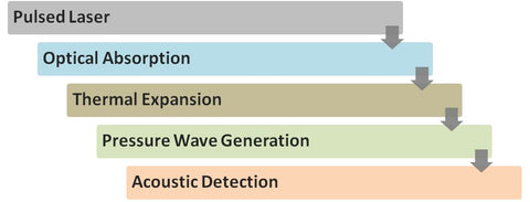
The photoacoustic effect was discovered by Alexander Graham Bell in the late 19th century and describes the event of acoustic waves generated by the absorption of light energy.1 Photoacoustic imaging works by using a pulsed laser to irradiate tissue. The light is absorbed by photoabsorbers naturally present in tissue or contrast agents added to tissue and this causes thermoelastic expansion which then translates through the tissue as a pressure or acoustic wave. The amount of thermal expansion determines how much pressure is generated. The acoustic waves are detected by an ultrasound transducer and translated into an image of the location of the endogenous or exogenous contrast agents.
The contrast in photoacoustic images depends primarily on how much light is absorbed by the tissue and/or the contrast agent. The optical absorption of different materials varies with wavelength, which makes it possible to identify and distinguish between different tissue components or exogenous contrast agents using photoacoustic imaging.
Advantages of Photoacoustic Imaging
Photoacoustic imaging is an excellent biomedical imaging diagnostic tool because it uses non-ionizing radiation to image tissue with high resolution and contrast in real time and at long penetration depths.3 While native tissue components, like hemoglobin and melanin, provide some photoacoustic contrast, the addition of exogenous agents opens an endless toolbox for molecular exploration of diseased tissue. The use of contrast agents in photoacoustic imaging also enables functional imaging which can be used to diagnose and characterize disease, and monitor the success of treatments.
Photoacoustic imaging when combined with ultrasound (USPA imaging) offers several advantages over other existing animal imaging modalities. Ultrasound imaging by itself is an excellent low-cost, non-ionizing portable imaging technology capable of morphologic imaging, but provides limited molecular contrast. In USPA imaging, the ultrasound is used primarily to visualize anatomical structures and photoacoustic imaging provides complementary information about the tissue composition and functionality.
Overall, photoacoustic imaging has several well known advantages:
- No harmful ionizing radiation
- Sub-millimeter structure image resolution with high penetration depth
- Near real-time imaging capability
- Excellent contrast agents and molecular targeting at imaging depth
- Requires only modest floor-space and offers ultra-mobile units for point of care use
- Greater convenience at a lower cost
Gold Nanoparticles for Photoacoustic Imaging
The use of contrast agents in photoacoustic imaging allows researchers to ascertain more information about the structure or function of living tissue. Our gold nanoparticles are specially designed for use as contrast agents in photoacoustic imaging.
Our gold nanorods are fine-tuned to exhibit localized surface plasmon resonances between 780 and 1064 nm, the near infrared optical window providing the highest differentiation from native tissue absorbers. Their silica coating enables efficient heat transfer from the particles, improves their thermodynamic stability, and enhances photoacoustic signal strength. Additionally, our upcoming antibody-conjugated line enables targeted imaging for tumors and other diseases.

References
- Bell AG. Upon the production of sound by radiant energy. American Journal of Science. 1880;20:305–324.
- Srivalleesha Mallidi, Geoffrey P. Luke, Stanislav Emelianov, Photoacoustic imaging in cancer detection, diagnosis, and treatment guidance, Trends in Biotechnology, Volume 29, Issue 5, May 2011, Pages 213-221, ISSN 0167-7799, http://dx.doi.org/10.1016/j.tibtech.2011.01.006.
- G. P. Luke, D. Yeager, and S. Y. Emelianov, "Biomedical Applications of Photoacoustic Imaging with Exogenous Contrast Agents," Annals of biomedical engineering, Nov 3 2011.