Contrast Agents
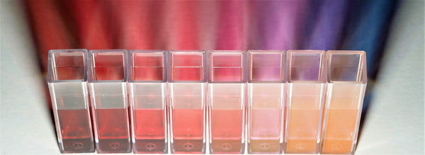
Premium Gold NanoSpheres - left to right 5nm, 10nm, 20nm, 40nm, 60nm, 80nm, 100nm, 120nm colloidal gold
Contrast agents can significantly improve the quality of a biomedical image and enable targeted or functional imaging. Optical contrast agents typically use phenomena such as scattering or absorption to enhance the signal depending on the imaging modality employed.
The use of contrast agents in imaging allows researchers to ascertain more information about the structure or function of living tissue. This information can help physicians make more educated decisions about appropriate treatment for their patients and also has the potential to allow monitoring of some therapies.
Why Use Exogenous Contrast Agents
Exogenous contrast agents such as dye or nanoparticles are utilized to improve the contrast of natural tissue and help identify pathological conditions.2 Exogenous optical contrast agents are designed to absorb light more strongly than local endogenous optical contrast that occurs naturally in tissue from melanin, hemoglobin, and fat.
In many cases, it is advantageous to differentiate the contrast from exogenous and endogenous absorbers. The graph below shows the absorption of various types of endogenous agents across a spectrum of wavelengths. There exists an “optical window” between ~700-1100 nm where the optical absorption of the endogenous tissues is minimized (shown in pink). Most exogenous contrast agents are designed to absorb light in this window in order to maximize their differentiation from endogenous absorbers and increase imaging depth. In some applications, hyper-spectral or multi-spectral imaging analysis techniques may be used to differentiate between endogenous absorbers and exogenous contrast agents.1,2
Exogenous contrast agents are extremely versatile since their optical absorption properties can be tailored to resonate at specific wavelengths in the optical window. Metallic nanoparticles are an excellent example since their surface plasmon resonance (SPR) can be easily tuned by varying their overall size and aspect ratio.
Contrast Agents in Optical and Photoacoustic Imaging
Optical imaging is an attractive biomedical imaging technique because it uses non-ionizing radiation and can be enhanced by the use of contrast agents. It is possible to differentiate between soft tissues using endogenous contrast, however when exogenous contrast agents are used the imaging possibilities are expanded to potentially imaging molecular events and cellular function.
As shown in Figure 1, in optical imaging, there is an “optical window” between 700-1100 nm where the optical absorbance of the endogenous tissues is minimized . This means that the near-infrared region is typically a target wavelength because the endogenous tissue does not interfere with the signal from the exogenous contrast agents.
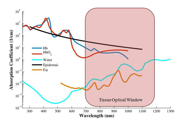
Figure 1: Optical Absorbance of Endogenous Tissues
Photoacoustic imaging, a type of optical imaging combined with ultrasound detection, is sensitive to the optical properties of the tissue as well, but it is less limited by light scattering because light travels in one direction and sound is monitored on return. It utilizes short (nanosecond) pulses of laser light usually in the same optical window mentioned above.3 Of the four mechanisms of photoacoustic signal generation, metallic contrast agents use thermal expansion.4 Contrast agents for photoacoustic imaging work by absorbing light of a certain wavelength, heating the water surrounding them causing it to thermoelastically expand. That expansion creates a pressure wave which is detected by the transducer. The amount of pressure generated depends on the properties of the contrast agent, the fluence of the light used, and parameters of the tissue being imaged.1 Using nanoparticle contrast agents with photoacoustic imaging is very powerful because the exogenous nanoparticle contrast agents can be targeted to a molecular biomarker of interest and their location in vivo can be visualized and differentiated from native endogenous absorbers like oxygenated and deoxygenated hemoglobin as shown in Figure 2.
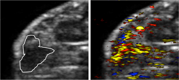
Figure 2: Ultrasound (left) and ultrasound and photoacoustic combined image (right) of a mouse model of pancreatic cancer. The cancer is outlined on the left and appears as a hypoechoic region. The photoacoustic overlay on the right shows the location of deoxygenated hemoglobin (blue), oxygenated hemoglobin (red) and nanoparticles (gold). Images are 17.5 mm by 12 mm.
Choosing The Right Contrast Agent
There are many considerations when choosing an appropriate contrast agent for a particular application. The ideal contrast agent depends on the imaging modality used and the specific application. The size, shape, surface chemistry, stability, and absorbance spectrum are all important parameters to take into account.It is also important to consider the environment in which you are imaging and what endogenous agents might be present.
Many contrast agents can be tailored for specific applications. For example, gold nanorods can be synthesized with varied aspect ratios to have different optical properties or they can be functionalized with silica coatings of different thicknesses to modify their plasmonic properties and increase the signal amplitude in photoacoustic imaging. If molecular targeting is desired for a particular application, the surface chemistry of the contrast agent is crucial since this will determine how the particle interacts with biology and the different strategies that can be employed for bioconjugation to antibodies or other targeting moieties.
Our Contrast Agents
The contrast agents produced by NanoHybrids offer many benefits:
- Our gold nanospheres are offered in three different sizes (5, 10, and 20 nm) to serve a variety of needs. The larger gold nanospheres will generate a greater photoacoustic signal. The size of the nanosphere will also affect the level of clearance.
- Our gold nanorods have full tunability to specific wavelengths (780-1064 nm) for use in many applications.
- Researchers also have the option of choosing silica coated nanoparticles to improve thermodynamic stability. In addition, the silica coating also enhances the photoacoustic response and enables multi-modal imaging.
- Our products are high quality and tested to ensure that the product you receive is up to the highest standard of monodispersity.
- Our contrast agents are suitable for many applications such as:
- Photoacoustic imaging
- Optical coherence tomography (OCT)
- Molecular tracking
- Monitoring treatment progress
- Spectroscopy
- Lateral flow assays
- Theranostics
Please feel free to contact us at info@nanohybrids.net if you need more information about which contrast agents would work best for your application.

References
- G. P. Luke, D. Yeager, and S. Y. Emelianov, "Biomedical Applications of Photoacoustic Imaging with Exogenous Contrast Agents," Annals of biomedical engineering, Nov 3 2011.
- "Optical Imaging." NIH Fact Sheets. National Institute of Health, Oct. 2010. Web. <http://report.nih.gov/nihfactsheets/ViewFactSheet.aspx?csid=105>.
- American National Standard for Safe Use of Lasers. Laser Institute of America; ANSI Z136.1 (2000).
- Emelianov, S. et al. Synergy of ultrasound, elasticity, and optoacoustic imaging for improved detection and differentiation of cancerous tissue. The Journal of the Acoustical Society of America 115, 2411 (2004).